flow cytometry results for lymphoma
They provide the theory and key practical aspects of flow cytometry enabling immunologists to avoid the common errors that often. These guidelines are a consensus work of a considerable number of members of the immunology and flow cytometry community.

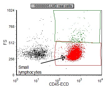
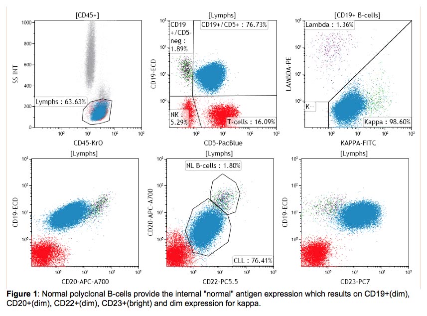
Flow Cytometric Presentation Of A Large B Cell Lymphoma A Forward Download Scientific Diagram
Images from quadrant Q2 positive for both stains acquired as a single event show the CAR T.

. Although rare Hodgkin lymphoma is one of the best-characterized malignancies of the lymphat ic system and one of the most readily curable forms of malignant disease. Results showed that in 916 56 percent the diagnosis of lymphoma or cancer could be suspected by flow cytometry alone while 416 were consistent with the final tissue diagnosis of normal or reactive hyperplasia. Flow cytometry is a methodology which is utilized during analysis of a heterogeneous population of cells according to different cell surface molecules size and volume which allows the investigation of individual cells-FACS together with flow cytometry can measure and characterize multiple cell generations by using highly specific antibodies tagged with.
The imaging capability lets you look more deeply into results to. A special technique called immunohistochemistry IHC is used to identify CD20 and determine whether an abnormal cancerous white blood cell lymphocyte in particular is a B. Three samples that came from patients who had morphologic evidence of malignant disease on biopsy two Hodgkins disease and one large cell lymphoma.
CD20 can be used to tell the difference between these two cancers in that test results for CD20 would usually be positive in the case of DLBCL but negative for ALCL. Built on more than 45 years of BD experience and leadership in flow cytometry and multicolor analysis the BD FACSCanto Flow Cytometry Systems deliver reliable performance accuracy and ease-of-use for todays busy clinical laboratories. Hodgkin lymphoma is a malignancy characterized histopathologically by the presence of Reed-Sternberg cells in the appropriate cellular background.
How Is It Tested. 2 The incidence rate has remained fairly steady over time it is. Lymphoma View All Blood Cancers.
In the figure engineered CAR T immunotherapy cells were co-incubated with Ramos lymphoma cells and stained acquired and imaged on the Attune CytPix Flow Cytometer. Get more information from the BD FACSCanto System brochure. Even show interactions between cells.

Summary Of Flow Cytometry Immunophenotypic Results For Anaplastic Large Download Table

Utility Of Flow Cytometry In Diagnosing Angioimmunoblastic T Cell Download Scientific Diagram

Diffuse Large B Cell Lymphoma Dlbcl Flow Cytometry

Follicular Lymphoma Fl Flow Cytometry

Flow Cytometry Results Flow Cytometric Graphs Showing Positivity For Download Scientific Diagram

Selected Flow Cytometric Immunophenotyping Plots From Fine Needle Download Scientific Diagram

International Clinical Cytometry Society

Flow Cytometric Immunophenotyping Performed On The Same Plasmablastic Download Scientific Diagram

Flow Cytometry Immunophenotypic Diagnosis Of B Cell Non Hodgkin Lymphomas On Fine Needle Aspirate Of Lymph Node Khanom Kh Tarafder S Sattar H Clin Cancer Investig J